
-
Publish Your Research/Review Articles in our High Quality Journal for just USD $99*+Taxes( *T&C Apply)
Offer Ends On
Publish Your Research/Review Articles in our High Quality Journal for just USD $99*+Taxes( *T&C Apply)
Offer Ends On
Philip L Millstein DMD, MS*
Corresponding Author: Philip L Millstein, Department of Restorative Dentistry, Harvard School of Dental Medicine, Boston, Massachusetts, USA.
Received: August 20, 2026 ; Revised: August 23, 2026 ; Accepted: August 25, 2026 ; Available Online: August 26, 2026
Citation: Millstein PL. (2026) Posterior Occlusal Stability Assures Anterior Occlusal Stability. Instability Assures Failure. J Oral Health Dent Res, 5(3): 1-3.
Copyrights: ©2026 Millstein PL. This is an open-access article distributed under the terms of the Creative Commons Attribution License, which permits unrestricted use, distribution, and reproduction in any medium, provided the original author and source are credited.
Views & Citations
Likes & Shares
Occlusal stability is essential for dental health. Posterior occlusal contact sustains anterior occlusal contact. Loss of posterior occlusal contact jeopardizes the working dentition. Periodic clinical examination is suggested.
Keywords: Occlusal contact, Holding cusps, Occlusal Stability, Instability.
The posterior occlusion evolves as teeth erupt. The dentition becomes stable with occlusal contact. A stable occlusion can remain functional over a lifetime; a failed occlusion will collapse (1). Periodic intra oral examinations should include the occlusion (2). Past findings should be compared with present findings however there are no records. Dentists are not trained to take occlusal records. An intra oral examination is incomplete without comparing occlusal records. The loss of occlusal contact stability leads to bite collapse which is a silent disease. Full mouth implant restorations or those in a mixed dentition present added challenges because implants exhibit little touch sensitivity and are immobile (3). A general practitioner can restore a broken tooth with a new occlusal surface but it is only a repair. In restorative dentistry we often use zirconia restorations because they always fit (4). They are strong, inexpensive and white. Internally they are constructed with a large cement space (300-500 microns) and externally they are under articulated (almost touching). There are no remakes. The restoration does not make occlusal contact with it’s antagonist. It does not support the posterior occlusion. A crown with a large cement space will shift it’s assigned occlusal position upon cementation compression. It twists (5). There are ordinarily eight posterior occlusal stops. Eliminate one with a repair and occlusal contact is lost. Six occlusal contact areas remain. Construct two crowns with uncertain occlusal contact areas and the posterior occlusion is reduced to four occlusal contact stops (6). The “bite” becomes unstable. Without stable posterior contact the occlusal forces will shift to the anterior (7). The anterior segment may have it’s own pattern of occlusion such as anterior guidance and cuspid rise (8). These movements are now negatively affected. The occlusal pathology that follows is unknown because the posterior occlusion is not stable. The existing occlusion self adjusts in a silent attempt to stabilize (9).
A practitioner upon oral examination should be aware of occlusal change but practitioner’s will rarely alter their daily routine to look for occlusal disharmony. Clinicians are not trained to evaluate the occlusion. Dentistry teaches caries control, oral pathology and orthodontia. It does not teach students about occlusal pathology or how to prevent it. People lose as many teeth from occlusal pathology as from caries infections. Dentists work with caries but they don’t work with the occlusion. In general they do not know what to do with occlusal pathology nor do their teachers. They talk about it. They prescribe a conventional bite guard after a loss of occlusal support. This is the treatment. There is no basic understanding of occlusal health. The bite slowly collapses as the dentist watches it collapse. Repair is now inevitable but what does a dentist do? Vertical dimension is lost. Diagnosis and treatment planning are required to restore an occlusal profile (10). A simple way to examine an occlusal scheme and to plan a treatment is to make an occlusal replica (11). This places the clinician in control. Treatment is planned extra orally. Ideas can be shared. To make an occlusal replica the clinician needs to take a closed intra oral occlusal impression in a triple tray using a non set silicone impression material. The impression is placed on an illuminated light box and photographed from above (12). The occlusal record is now permanently recorded and can be viewed on screen. A non set viscous silicone material records the occlusal contacts and near occlusal contact areas along with guided excursions (13).
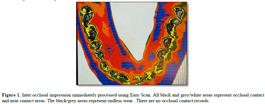
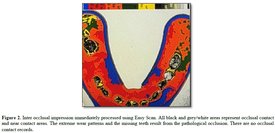
A permanent set type of silicone impression material can be used but it is more expensive and time consuming. It does not record excursions. Image J freeware (Easy Scan) is used to numerically evaluate the black and white photographic images.
The numbers can be converted to colors for easy viewing. Figure. 1, 2. Comparisons and evaluations can be made. New designs can be implemented. All clinical evaluation and planning is done extra orally. Recorded images are saved (14).
 Occlusal health is essential to good health. The only person who can judge the working occlusion and if needed treat it is the attending dentist. The only non commercial places to learn about the occlusion is in a dental school or study club. Occlusal pathology like caries pathology requires research and development. Clinical research is essential. Prevention is the goal.

No Files Found


Internationally Accepted
Share Your Publication :