
-
Publish Your Research/Review Articles in our High Quality Journal for just USD $99*+Taxes( *T&C Apply)
Offer Ends On
Publish Your Research/Review Articles in our High Quality Journal for just USD $99*+Taxes( *T&C Apply)
Offer Ends On
Mohamad El-Moheb, Haseeb Al Dary, Lina Droubi, Moath Omar Badawi and Bruno Viana Reis*
Corresponding Author: Bruno Viana Reis, Unique Dental, 90-97 Cork Street, Dublin, Ireland.
Received: Unique Dental ; Revised: August 02, 2026 ; Accepted: August 04, 2026 ; Available Online: August 06, 2026
Citation: El-Moheb M, Al Dary H, Droubi L, Badawi MO & Reis BV. (2026) Elemental Aluminum in Bovine Implant-Bed Bone Following Dental Implant Placement: An Exploratory Ex Vivo Study. J Oral Health Dent Res, 5(3): 1-11. https://doi.org/10.5281/zenodo.21918396
Copyrights: ©2026 El-Moheb M, Al Dary H, Droubi L, Badawi MO & Reis BV. This is an open-access article distributed under the terms of the Creative Commons Attribution License, which permits unrestricted use, distribution, and reproduction in any medium, provided the original author and source are credited.
Views & Citations
Likes & Shares
Statement of problem. Mechanical interaction between a dental implant and the osteotomy walls may alter the implant surface and transfer metallic material to the surrounding bone. Aluminum may originate from aluminum-containing titanium alloys, residual aluminum oxide used during surface blasting, or other manufacturing and procedural sources.
Purpose. The purpose of this exploratory study was to determine whether elemental aluminum could be detected in implant-bed bone following dental implant placement.
Material and methods. Seven commercially available implant systems were placed in dense bovine rib bone according to the drilling protocols and insertion-torque limits recommended by their respective manufacturers. Three untreated portions of the same rib were collected as negative biological controls. Implant removal was attempted using counter-torque. Five implants were successfully retrieved, whereas two were excluded after failure of the implant–driver hexagonal engagement because their removal would have required trephination or bone sectioning. The untreated controls and recovered implant-bed specimens underwent microwave digestion followed by inductively coupled plasma optical emission spectrometry. Aluminum concentrations were expressed in micrograms per gram of bone. Results reported as 0.00 µg/g were classified as not detected because no aluminum signal had been identified. [23]
Results. No aluminum signal was detected in any of the three untreated bone controls. Aluminum was detected in four of the five analyzable implant-bed specimens, at concentrations ranging from 0.042 to 0.140 µg/g. The highest concentrations were measured in the ROOTT R and Biotech Kontact specimens, at 0.140 and 0.120 µg/g, respectively. Lower concentrations were detected in the Straumann SLA Active Ti-Zr and ISY Camlog specimens, at 0.065 and 0.042 µg/g, respectively. No aluminum signal was detected in the DXL Smart implant-bed specimen. Because each implant system was represented by a single specimen, no inferential statistical comparisons were performed. [1-4]
Conclusions. Elemental aluminum was detected in four of five implant-bed specimens but not in untreated bone controls, supporting the hypothesis that the sequence of osteotomy preparation and implant placement may introduce aluminum into recipient bone. Aluminum-containing alloys and aluminum oxide-associated surface treatments represent plausible sources, although definitive attribution was not possible. The detected quantities cannot be interpreted as evidence of toxicity, systemic exposure, or neurodegenerative risk. Replicated studies incorporating drilling-only controls, recorded insertion torque, non-destructive specimen retrieval, validated analytical sensitivity, and complementary SEM–EDS characterization are required.
Keywords: aluminum; implant-bed bone; metallic debris; particle release; implant surface.
CLINICAL IMPLICATIONS
Trace elemental aluminum may be transferred to implant-bed bone during osteotomy preparation and implant placement, even when implants are placed within manufacturer-recommended torque limits. Because this exploratory study used one specimen per implant system and did not determine chemical form or biological effects, the findings should not alter clinical practice but support further controlled investigation of implant alloys and surface-treatment residues. [1-4,15,16]
INTRODUCTION
Titanium and titanium alloys are widely used for endosseous dental implants because of their favorable mechanical properties, corrosion resistance, biocompatibility, and capacity to support osseointegration. Contemporary implant systems differ substantially from the original machined titanium designs, particularly in their alloy composition, macrogeometry, and surface microtopography. Roughened surfaces produced by blasting, acid etching, anodization, or combined treatments are intended to increase bone–implant contact and accelerate biological fixation. At placement, however, the implant is inserted into an osteotomy that is generally narrower than the maximum external diameter of its threads. The resulting compressive, frictional, and shear forces contribute to primary mechanical stability but may also alter the surface that was manufactured to interact with the surrounding bone. [10,11]
Experimental evidence demonstrates that implant insertion can reduce surface roughness and detach material from the implant surface. Senna et al. reported surface damage and loose titanium and aluminum particles at the bone–implant interface after implants were placed in fresh bovine rib, with debris concentrated predominantly in the crestal cortical region.1 Barrak et al. subsequently demonstrated the immediate release of metallic particles and ions after placement of different implant systems and found that the quantity released was influenced by implant material and design.2 More recently, Kheder et al. confirmed the presence of titanium particles embedded within bovine implant beds and associated particle release with changes in implant surface roughness. [1-3]
Dodo et al. further examined this phenomenon in 2025 by comparing implant surfaces before and after insertion into fresh bovine rib under a high-insertion-torque protocol. Surface damage was concentrated at the crests of the implant threads, while the thread valleys showed comparatively little alteration. Loose titanium particles ranging from nanometres to micrometres were identified by scanning electron microscopy and energy-dispersive X-ray spectroscopy, particularly within the crestal cortical bone. These findings indicate that insertion-related material release is not necessarily distributed uniformly along the implant surface but occurs preferentially where thread features experience the greatest mechanical interaction with bone. [4]
Aluminum is of particular interest because it may be associated with dental implants through more than one pathway. It is an alloying element in Ti-6Al-4V and related titanium alloys, where it contributes to mechanical strength, while aluminum oxide particles may also be used as the abrasive medium during surface-blasting procedures. Aluminum detected in implant-bed bone could therefore originate from abrasion of an aluminum-containing implant alloy, residual alumina associated with surface processing, manufacturing contamination, or a combination of these sources. The detection of both titanium and aluminum debris at the bone–implant interface in previous experimental work supports the plausibility of insertion-related transfer, but the precise origin, chemical form, and biological availability of the detected aluminum cannot be assumed from elemental analysis alone. [17,18,19,20]
The biological significance of aluminum depends not only on its total quantity but also on its chemical form, particle size, solubility, localization, persistence, and capacity for cellular uptake or systemic distribution. Aluminum exposure has been investigated in relation to cytotoxicity and neurological and neurodegenerative disorders, including Alzheimer’s disease, although epidemiological findings remain inconsistent and a direct causal relationship has not been established.5-8 A prospective Canadian cohort, for example, did not demonstrate a statistically significant overall association between aluminum concentrations in drinking water and incident Alzheimer’s disease, although findings in genetically susceptible subgroups supported the need for continued investigation. [5,6,21,22]
The quantities detected locally after implant placement cannot therefore be directly related to neurodegenerative disease or interpreted as evidence of systemic toxicity. Nevertheless, exposure to aluminum may arise from multiple environmental, dietary, occupational, and medical sources. If aluminum introduced into peri-implant bone is subsequently solubilised, mobilized, or systemically distributed, it could theoretically represent an additional contribution to cumulative exposure. From a biomaterials and precautionary perspective, identifying and reducing potentially avoidable sources of metallic residues may be desirable, provided that this can be achieved without compromising implant strength, surface performance, primary stability, or osseointegration. Determining whether aluminum is transferred to the recipient bone during implant insertion is therefore relevant even when the clinical significance of the detected quantity remains unknown. [27,28,30]
Although previous studies have characterized insertion-related surface damage and identified metallic particles using microscopy and elemental mapping, quantitative information regarding total aluminum in the recipient bone remains limited. Accordingly, the objective of this exploratory ex vivo study was to determine whether elemental aluminum could be detected in implant-bed bone following the placement and removal of commercially available dental implants with different reported alloy compositions and surface treatments. Three untreated specimens from the same bovine rib were analyzed as negative biological controls, and total aluminum concentrations were determined by inductively coupled plasma optical emission spectrometry. [1-4]
It was hypothesised that no aluminum signal would be detected in untreated bone controls, whereas aluminum would be detectable in at least some implant-bed specimens. The study was also intended to generate preliminary hypotheses concerning the possible contributions of implant alloy composition and surface treatment. Because the analytical method measured total elemental aluminum after complete digestion of the bone specimens, it was not designed to determine whether the aluminum was present as metallic fragments, aluminum ions, aluminum oxide particles, or another chemical species, nor to establish its definitive source or biological fate. The null hypothesis was that no aluminum signal would be detected in either untreated controls or implant-bed specimens. [24]
MATERIAL AND METHODS
Study design and experimental substrate
This exploratory ex vivo study was designed to determine whether elemental aluminum could be detected in bone after the placement and subsequent removal of commercially available dental implants. Seven implant systems were initially included, with one implant representing each system. A bovine rib segment was used as the experimental substrate because of its high density and was macroscopically considered comparable to D1-type bone. Before any osteotomy preparation or implant insertion, three separate specimens were collected from untreated regions of the same rib. These specimens constituted the negative biological control group and underwent the same preparation, digestion, and elemental analysis as the implant-bed specimens. Although these specimens were identified as “Blank 1,” “Blank 2,” and “Blank 3” in the original laboratory report, they are referred to throughout the present study as untreated bone controls because they contained biological tissue and were not analytical reagent blanks. [24]
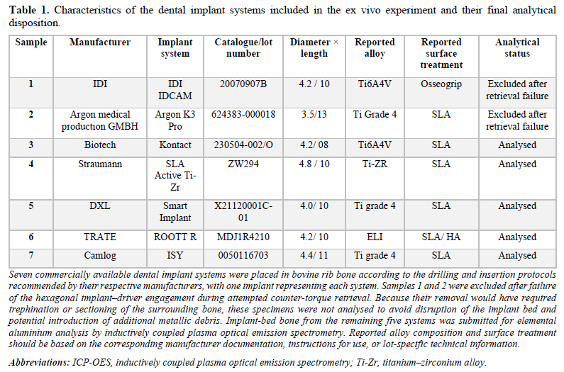
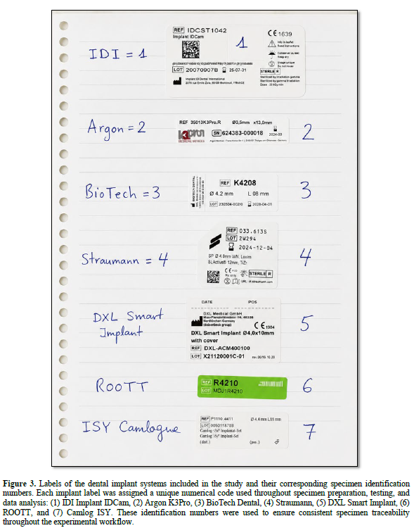
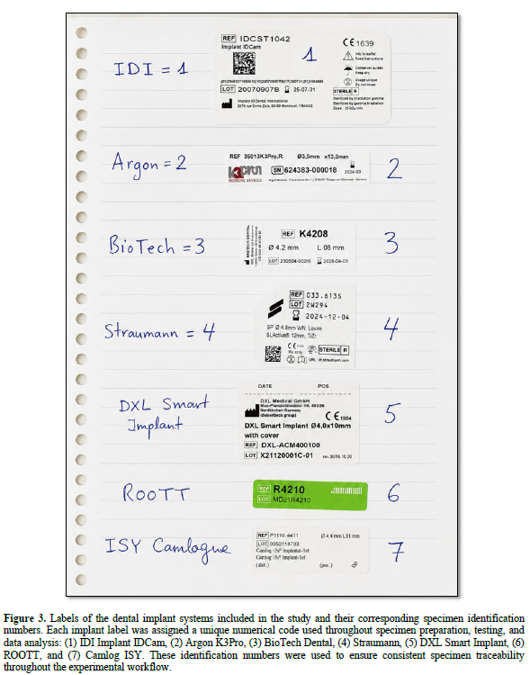
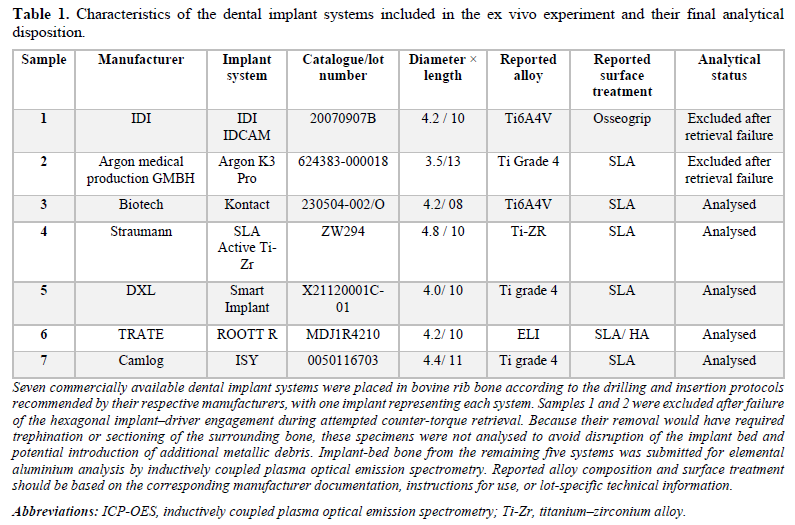
The seven implant systems evaluated were IDI IDCAM, Argon K3 Pro, Biotech Kontact, Straumann SLA Active Ti-Zr, DXL Smart Implant, ROOTT R, and ISY Camlog. The specific manufacturer, commercial designation, catalogue or lot number, implant dimensions, reported alloy composition, and surface treatment of each system are presented in Table 1. Each implant system was represented by a single specimen; consequently, the investigation was designed as an exploratory proof-of-concept study rather than as a statistical comparison of implant brands, materials, or surface treatments. [17,18] (Figure. 3)
Osteotomy preparation and implant insertion
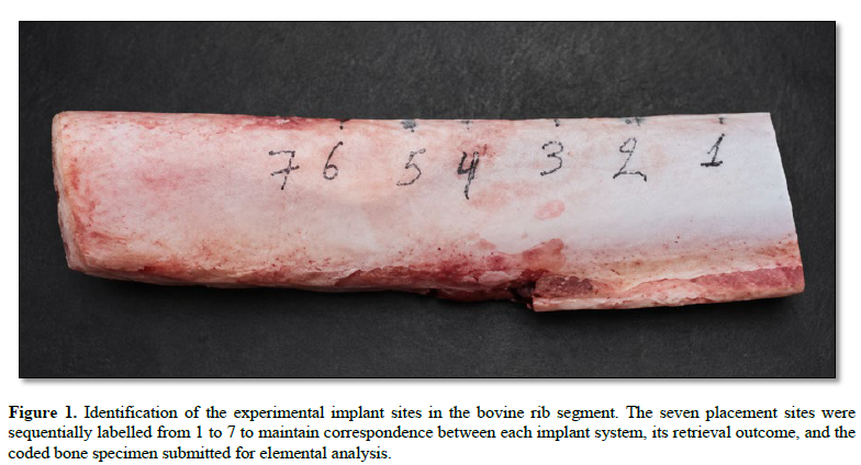
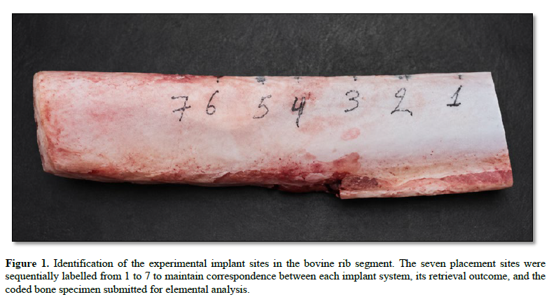
A separate osteotomy was prepared for each implant using the drilling sequence recommended by the corresponding manufacturer. Implant placement was performed in accordance with the manufacturer’s surgical protocol and within the maximum insertion-torque limit specified for each system. The implants were inserted into distinct regions of the bovine rib, avoiding overlap between adjacent osteotomies. The use of dense bone created a high-friction environment in which mechanical interaction between the implant threads and the osteotomy walls could occur during insertion. The sequentially labeled experimental sites are shown in Figure 1 (Fig. 1). [15,16]
The implant diameter and length, final drill diameter, osteotomy protocol, insertion speed, irrigation conditions, maximum insertion torque, and any use of tapping or countersinking are reported in Table 1 or should be added where these data were recorded. When exact numerical insertion-torque values were not documented, the procedure was described only as having been performed within the torque limits recommended by the respective manufacturers, without retrospective estimation of numerical values. [15,16]
Implant retrieval and specimen eligibility
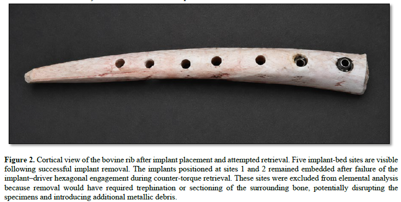
After insertion, removal of each implant was attempted by applying counter-torque through the corresponding implant driver. Five implants were successfully retrieved, allowing collection of the associated implant-bed bone for elemental analysis. The IDI IDCAM and Argon K3 Pro implants could not be removed by conventional counter-torque. During attempted reverse rotation, the hexagonal engagement between the implant and the driver failed, causing the driver to rotate without transmitting sufficient counter-torque to the implant. Both implants therefore remained mechanically engaged within the bone. The cortical view after attempted retrieval is shown in Figure 2 (Fig. 2). [1-4]
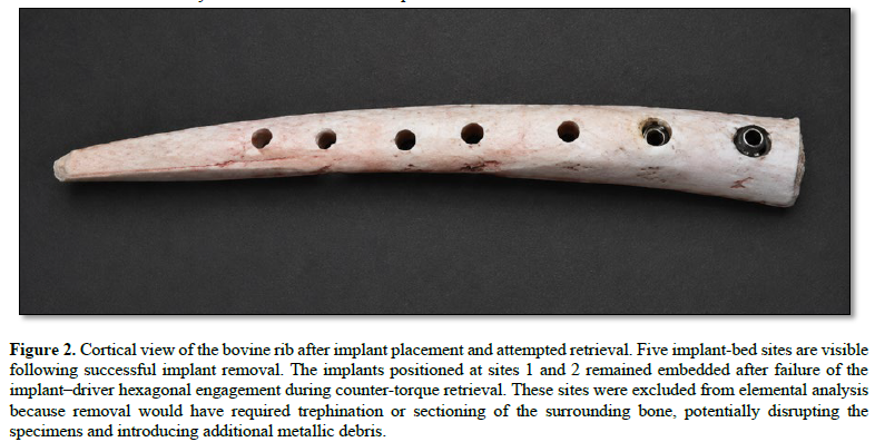
Retrieval of these implants would have required the use of a trephine bur or physical sectioning of the surrounding bone. These procedures were not performed because they could have introduced additional metallic debris, removed part of the implant-bed tissue, altered the distribution of material surrounding the implant, or produced specimens that were not directly comparable with those obtained after conventional implant removal. The two sites were therefore classified as retrieval-related technical exclusions and were not submitted for chemical analysis. The exclusions resulted from failure of the driver–implant engagement during retrieval and not from exceeding the manufacturers’ recommended insertion-torque limits. Accordingly, seven implants were placed, seven retrieval procedures were attempted, and five implant-bed specimens were available for analysis. [1-4]
Specimen collection and blinding
Following successful implant removal, bone associated with each implant bed was collected and prepared for analysis. The precise dimensions and method used to isolate the implant-bed specimens should be reported if documented in the original experimental records. The three untreated bone controls and the five implant-bed specimens were assigned numerical codes before being sent to the analytical laboratory. The laboratory was not informed of the implant brands, alloy compositions, surface treatments, or experimental conditions associated with the coded specimens. The allocation key was retained separately and the sample identities were disclosed only after completion of the elemental analysis. [23]
Reagents and calibration standards
All solutions used for specimen digestion, calibration, and instrument rinsing were prepared using ultrapure water. Nitric acid at a concentration of 69% and hydrochloric acid at a concentration of 35% were obtained from Merck. Undiluted nitric acid was used for microwave digestion, while the calibration solutions were prepared in 1% nitric acid using a 1000-ppm single-element aluminum standard supplied by Merck. Two rinsing solutions were used during the inductively coupled plasma analysis. The first contained 5% nitric acid and 1% hydrochloric acid, whereas the second contained 1% nitric acid and corresponded to the acid matrix of the diluted digestion samples. The concentrations of the individual calibration standards and the complete calibration range should be reported from the original laboratory analytical record. [23]
Microwave digestion
A weighed portion of bone from each untreated control and implant-bed specimen was transferred to a microwave digestion vessel and combined with undiluted nitric acid. Digestion was performed using a CEM Mars 5 microwave system. CEM XP-1500 vessels with a nominal capacity of 50 mL and CEM Xpress vessels with nominal capacities of 25 or 75 mL were used together with polytetrafluoroethylene magnetic stirrers. After the bone specimen and nitric acid had been added, the samples underwent a 20-minute predigestion period. [23]
The microwave program comprised a 20-minute temperature ramp to 180°C, followed by a 25-minute holding period at the same temperature. A power setting of 1200 W was used when more than six vessels were processed simultaneously on the microwave turntable. Magnetic stirring was maintained at the laboratory’s medium setting during digestion. After completion of the program, the vessels were allowed to cool to room temperature before being opened. The initial mass of each bone specimen, the volume of nitric acid added, the final digestion volume, and any dilution performed before instrumental analysis should be inserted from the original laboratory worksheets. [23]
Elemental aluminum analysis
The digested specimens were analyzed for elemental aluminum using inductively coupled plasma optical emission spectrometry. The instrument was identified in the original report as a Shimadzu ICP-9000; the exact model designation should be confirmed with the analytical laboratory before final submission. The reported operating conditions included a radio-frequency power of 1.2 kW, a cooling-gas flow of 10 L/min, a plasma-gas flow of 0.6 L/min, a carrier-gas flow of 0.7 L/min, and a sample aspiration rate of 0.6 mL/min. Sample introduction was performed using a coaxial nebuliser and a cyclone spray chamber, with axial or radial viewing according to the analytical configuration used for aluminum. [23]
The aluminum emission wavelength, calibration equation, calibration intercept, background-correction procedure, number of replicate readings, method detection limit, method quantification limit, calibration-verification procedure, analytical recovery, and measurement uncertainty should be added from the complete laboratory report when available. The analytical method quantified total elemental aluminum following complete digestion of the bone specimens. It did not determine whether the detected aluminum was present as metallic fragments, dissolved ions, aluminum oxide particles, alloy-derived debris, or another chemical species, and it did not independently establish the source of the aluminum. [23]
Definition and reporting of nondetected results
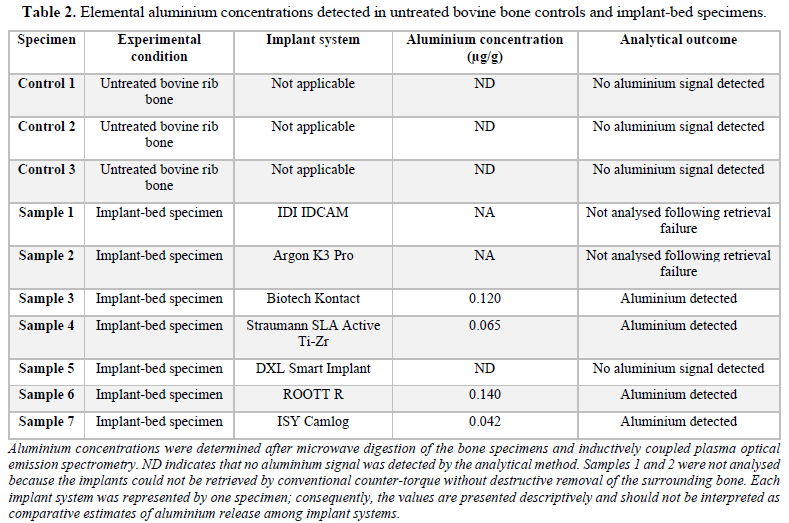
Aluminum concentrations were expressed in micrograms per gram of bone. Values reported by the laboratory as 0.00 µg/g indicated that no aluminum signal was detected during the instrumental analysis. These observations were therefore classified and reported as not detected rather than as an absolute aluminum concentration of zero. In the manuscript and results table, the abbreviation “ND” indicates that no aluminum signal was detected by the analytical method. The three untreated bone controls and the DXL Smart implant-bed specimen were classified as ND according to this reporting convention. [23]
Data analysis
The findings were evaluated descriptively. Because each implant system was represented by a single experimental specimen, no inferential statistical tests were performed and no statistical comparisons were made among implant brands, alloy compositions, or surface treatments. The two implant sites that could not be recovered without destructive intervention were treated as technical exclusions and were not assigned aluminum values. Results classified as ND were retained as non-detections and were not interpreted as quantitatively measured zero concentrations. [24]
Individual aluminum concentrations were reported for each of the five analyzable implant-bed specimens together with the findings from the three untreated bone controls. The study was intended to establish whether aluminum could be detected in implant-bed bone after implant placement and removal and to generate hypotheses regarding possible contributions from implant alloy composition, surface treatment, manufacturing residues, and insertion-related surface alteration. It was not designed to determine differences in aluminum release among commercial implant systems or to establish the biological or clinical significance of the detected concentrations. [1-4]
RESULTS
Seven dental implant systems were initially included in the experiment. All implants were placed in bovine rib bone according to the drilling and insertion protocols recommended by their respective manufacturers and within the corresponding insertion-torque limits. Five implants were successfully retrieved by counter-torque and their implant-bed specimens were submitted for elemental aluminum analysis. The IDI IDCAM and Argon K3 Pro implants could not be removed because the hexagonal implant–driver engagement failed during attempted reverse rotation, causing the driver to rotate without transmitting sufficient counter-torque to the implants. Because retrieval would have required trephination or sectioning of the surrounding bone, these two sites were excluded to avoid disruption of the implant bed and possible introduction of additional metallic debris. Accordingly, five implant-bed specimens and three untreated bone controls were available for analysis. [1-4]
No aluminum signal was detected in any of the three untreated bone controls. Among the five analyzable implant-bed specimens, aluminum was detected in four, with measured concentrations ranging from 0.042 to 0.140 µg/g. No aluminum signal was detected in the specimen associated with the DXL Smart implant. [1-4]
The highest aluminum concentration was measured in the implant bed associated with the ROOTT R implant, at 0.140 µg/g. The Biotech Kontact implant-bed specimen showed a concentration of 0.120 µg/g, followed by the Straumann SLA Active Ti-Zr specimen at 0.065 µg/g and the ISY Camlog specimen at 0.042 µg/g. The DXL Smart implant-bed specimen was reported by the laboratory as 0.00 µg/g, indicating that no aluminum signal was detected. Individual specimen results are presented in Table 2. [1-4]
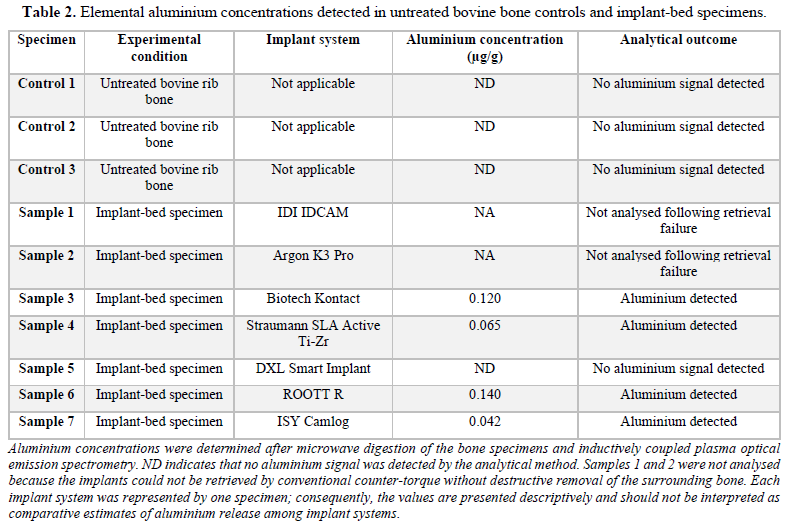
The two highest measured concentrations occurred in specimens associated with implants reported to contain aluminum within their titanium alloy composition. Aluminum was also detected in the implant beds associated with two implants reported to be manufactured from aluminum-free titanium or titanium–zirconium materials but treated using aluminum oxide blasting. Conversely, no aluminum signal was detected in one implant-bed specimen associated with a reportedly aluminum-free implant that had also undergone aluminum oxide surface treatment. These observations were recorded descriptively and were not interpreted as demonstrating a direct relationship between alloy composition, surface treatment, and the quantity of aluminum detected. [17,18,19,20]
Because each implant system was represented by a single specimen, no inferential statistical analysis or formal comparison among implant systems was performed. The findings therefore represent individual experimental observations rather than estimates of the average aluminum release associated with each implant system. [24]
Overall, elemental aluminum was detected in four of the five implant-bed specimens successfully recovered after implant placement, whereas no aluminum signal was detected in the three untreated bone controls. The detected concentrations varied among the individual implant systems, but the experimental design did not permit statistical comparison or definitive attribution of the aluminum to the implant alloy, surface treatment, or another procedural source. [1-4]
DISCUSSION
The null hypothesis was rejected because elemental aluminum was detected in four of the five analyzable implant-bed specimens, whereas no aluminum signal was detected in any of the three untreated bone controls. The measured concentrations ranged from 0.042 to 0.140 µg/g. The absence of a detectable signal in the untreated portions of the same bovine rib supports an association between the detected aluminum and the experimental sequence comprising osteotomy preparation, implant insertion, implant retrieval, and specimen processing. Nevertheless, the study design does not permit the aluminum to be attributed definitively to a specific stage of that sequence or to a particular component of the implant.
These findings are consistent with previous evidence that mechanical interaction between an implant surface and the osteotomy walls can modify the implant surface and transfer material to the surrounding bone. Senna et al. demonstrated surface damage and the presence of loose particles after dental implants were inserted into bone according to the recommended surgical protocol.1 Barrak et al. subsequently identified immediate particle and ion release from different implant systems and reported that the magnitude of release was influenced by implant material and design.[2] More recently, Dodo et al. showed that high insertion torque produced surface alterations concentrated particularly at the thread crests, with titanium particles identified in the adjacent cortical bone.[4] Together, these studies support the mechanical plausibility that frictional and shear forces generated during placement may detach material from the implant surface or dislodge residues retained from its manufacturing and surface-treatment processes.
An important distinction is that all implants in the present investigation were placed within the insertion-torque limits indicated by their respective manufacturers. The findings therefore suggest that detectable elemental transfer may occur even when the recommended surgical protocol is followed and does not necessarily require deliberately excessive insertion torque. This interpretation is compatible with the earlier findings of Senna et al., whereas Dodo et al. demonstrate how greater mechanical loading may intensify the same general phenomenon. However, because exact insertion-torque values were not available for all specimens in the present study, no relationship between torque and aluminum concentration can be established. Differences among the individual results may also have been influenced by implant diameter, thread design, taper, osteotomy dimensions, cortical thickness, surface roughness, and the contact area developed during placement. [23,24]
The distribution of the results raises two plausible, although unconfirmed, pathways for aluminum transfer. The two highest concentrations, 0.140 and 0.120 µg/g, occurred in implant-bed specimens associated with implants reported to contain aluminum as part of their titanium alloy composition. In these cases, abrasion of the implant alloy during insertion could theoretically have contributed to the detected aluminum. Aluminum was also detected at lower concentrations in the implant beds associated with the Straumann Ti-Zr and ISY Camlog implants, which were reported to be manufactured from nominally aluminum-free materials but to have surfaces produced using aluminum oxide blasting. In those specimens, residual alumina from the surface-treatment process represents a possible source. These interpretations remain hypotheses because the reported alloy compositions and surface treatments require confirmation from the relevant manufacturer documentation and because elemental analysis alone cannot identify the origin of the aluminum. [23,24]
The absence of a detectable aluminum signal in the DXL Smart implant-bed specimen is also relevant. That implant was reported to be manufactured from commercially pure titanium and treated using aluminum oxide blasting, yet no aluminum signal was detected. This observation indicates that alumina blasting does not inevitably result in detectable aluminum transfer under the conditions used in this study. Differences in post-blasting cleaning, surface decontamination, blasting-particle retention, surface roughness, implant macrogeometry, or local mechanical engagement may influence whether aluminum is transferred to the recipient bone. However, because only one specimen represented each implant system, the non-detection cannot be interpreted as evidence that the DXL system consistently releases no aluminum or that its manufacturing process is superior to those of the other systems. [19,20]
The three untreated bone controls strengthen the internal interpretation of the study. Because no aluminum signal was detected in any of these specimens, the positive findings in four implant beds are unlikely to be explained solely by the pre-existing aluminum content of the specific rib sections analyzed. Nevertheless, the untreated controls do not isolate the individual contribution of osteotomy preparation from that of implant insertion. A drilling-only procedural control, in which the complete osteotomy sequence is performed without implant placement, would be necessary to determine whether any detectable aluminum was introduced by the drilling procedure, surgical instrumentation, irrigation, sample handling, or another part of the experimental process. Future investigations should therefore include untreated bone controls, drilling-only controls, and implant-insertion groups processed simultaneously under identical analytical conditions. [24]
The analytical method is another central consideration when interpreting the results. Microwave digestion followed by inductively coupled plasma optical emission spectrometry quantified the total elemental aluminum present in each digested bone specimen. It did not establish whether the aluminum was present as metallic fragments, aluminum ions, aluminum oxide particles, alloy-derived debris, or another aluminum-containing compound. It also did not provide information about particle number, size, morphology, spatial distribution, or localization in relation to the cortical and cancellous portions of the osteotomy. Consequently, the present findings should be described as the detection of elemental aluminum in implant-bed bone rather than as the direct identification of aluminum particles. [23]
This distinction explains why the current findings complement rather than reproduce those of Dodo et al. Their use of scanning electron microscopy and energy-dispersive X-ray spectroscopy allowed surface alterations and individual metallic particles to be localized and chemically characterized. By contrast, the present method was capable of detecting and quantifying the total aluminum content of the digested bone specimen but sacrificed spatial and morphological information. A combined approach would therefore be preferable in future studies. ICP-OES or inductively coupled plasma mass spectrometry could quantify total elemental release, while scanning electron microscopy with elemental mapping could determine whether the aluminum occurs in discrete particles, establish its association with other elements, and localise it within the implant bed. [1-4]
The biological significance of the measured concentrations remains unknown. The present study did not evaluate cell viability, inflammatory signaling, osteoblast differentiation, bone formation, systemic absorption, or clinical outcomes. Experimental evidence has shown that ionic constituents associated with Ti-6Al-4V can affect aspects of osteogenic-cell differentiation under specific in vitro exposure conditions, including inhibition of osteocalcin synthesis. [9] Such findings establish biological plausibility but cannot be directly extrapolated to the much smaller quantities detected locally in the present ex vivo bone specimens. The chemical form, concentration, exposure duration, cellular availability, and experimental environment differ substantially.
Similarly, the aluminum concentrations reported here cannot be connected directly to Alzheimer’s disease or any other neurological or neurodegenerative condition. The literature has investigated aluminum exposure as a possible contributing factor in neurodegenerative disease, but the present experiment provides no evidence regarding systemic dissemination or neurological effects. Nevertheless, aluminum exposure may arise from multiple dietary, environmental, occupational, pharmaceutical, and medical sources. If aluminum transferred during implant insertion is subsequently dissolved, mobilized from the peri-implant tissues, distributed systemically, and retained, it could theoretically constitute an additional contribution to cumulative exposure. The present study did not investigate any of these stages, and the detected quantities must not be interpreted as establishing a measurable systemic burden. [5-7,29,31]
The significance of this cumulative-exposure hypothesis is therefore precautionary rather than causal. Even where an individual source is quantitatively small, identifying potentially avoidable exposure may remain relevant when the source provides no intended biological benefit. From a biomaterials and manufacturing perspective, unnecessary aluminum residues could potentially be reduced through alloy selection, optimization of blasting procedures, improved post-treatment cleaning, or the use of alternative surface-processing methods. Such changes would only be justified if they preserve the mechanical properties, surface characteristics, primary stability, and osseointegration performance required of the implant. The present results do not demonstrate that aluminum-free alloys or non-alumina surface treatments are clinically superior; they identify a question requiring controlled investigation. [23,24]
Two of the seven initially selected implant systems could not be included in the chemical analysis. Although both implants had been inserted within the torque limits indicated by their manufacturers, the hexagonal implant–driver engagement failed during attempted counter-torque retrieval. The driver consequently rotated without transmitting sufficient reverse torque, and removal would have required trephination or cutting of the surrounding bone. These destructive retrieval procedures were avoided because they could have introduced additional metallic debris, removed part of the implant bed, or redistributed material within the specimen. The exclusions were therefore methodologically justified, but they reduced the number of analyzable systems and may have introduced selection bias because the aluminum concentrations associated with those two implants remain unknown. No conclusion regarding the mechanical reliability of either implant connection can be drawn from one retrieval event per system.
The principal limitation of the investigation was the use of only one implant and one implant-bed specimen for each commercial system. The findings represent individual observations and cannot establish a reproducible aluminum-release profile for any manufacturer, alloy, or surface treatment. No statistical comparison or brand ranking is therefore appropriate. The use of bovine rib also limits direct extrapolation to clinical placement because human bone varies in cortical thickness, density, trabecular architecture, vascularity, and biological response. In addition, the absence of recorded insertion-torque values for each specimen, drilling-only controls, particle-level characterization, and complete analytical sensitivity data restricts the identification of the mechanisms responsible for the observed results. [23,24]
Future studies should use multiple independent specimens for each experimental condition and standardize implant dimensions, osteotomy undersizing, bone density, cortical thickness, insertion speed, irrigation, and recorded insertion torque. The protocol should include untreated bone, drilling-only, and implant-insertion controls. Implant retrieval should be avoided where possible by designing bone blocks that can be separated after insertion without unscrewing the implant, thereby preserving the interface and reducing the possibility of retrieval-related contamination. Total elemental analysis should be combined with surface profilometry, scanning electron microscopy, and elemental mapping. Comparing unused implants, retrieved implants, and implant-bed bone would help determine whether detected aluminum originated from alloy abrasion, retained alumina, manufacturing contamination, or another source. [1-4,24]
Within the limitations of this exploratory study, the absence of a detectable aluminum signal in untreated bone and its detection in four of five analyzable implant-bed specimens support the hypothesis that dental implant placement may introduce elemental aluminum into the recipient bone. The pattern of findings suggests that both aluminum-containing alloys and alumina-associated surface treatments may represent possible sources, although definitive attribution was not possible. The detected concentrations cannot be interpreted as evidence of local toxicity, systemic exposure, or neurological disease. They identify a potentially avoidable source of aluminum exposure and provide a rationale for replicated studies investigating its origin, chemical form, biological fate, and clinical relevance. [1-4,31]
CONCLUSIONS
Within the limitations of this exploratory ex vivo study, elemental aluminum was detected in four of the five analyzable implant-bed specimens, whereas no aluminum signal was detected in any of the three untreated bovine bone controls. The measured concentrations ranged from 0.042 to 0.140 µg/g. These findings support the hypothesis that the sequence of osteotomy preparation and implant placement may introduce aluminum into the recipient bone, even when implants are inserted according to the manufacturers’ recommended protocols and within their stated torque limits.
The observed pattern suggests that aluminum-containing titanium alloys and aluminum oxide-based surface treatments may both represent possible sources. However, because each implant system was represented by only one specimen and the analytical method quantified total elemental aluminum after complete digestion, the origin, chemical form, particle characteristics, and biological fate of the detected aluminum could not be determined. The results cannot be interpreted as evidence of local toxicity, systemic exposure, or a relationship with neurodegenerative disease. Nevertheless, implant-associated aluminum may represent an additional and potentially avoidable source of cumulative exposure. Replicated studies incorporating drilling-only controls, recorded insertion torque, non-destructive specimen retrieval, validated detection limits, and complementary SEM–EDS analysis are required to determine the source, distribution, bioavailability, and clinical significance of aluminum detected after dental implant placement. [23,24]
No Files Found


Internationally Accepted
Share Your Publication :