
-
Publish Your Research/Review Articles in our High Quality Journal for just USD $99*+Taxes( *T&C Apply)
Offer Ends On
Publish Your Research/Review Articles in our High Quality Journal for just USD $99*+Taxes( *T&C Apply)
Offer Ends On
Philip L Millstein DMD, MS*
Corresponding Author: Philip L Millstein, Department of Restorative Dentistry, Harvard School of Dental Medicine, Boston, Massachusetts, USA.
Received: August 02, 2026 ; Revised: August 04, 2026 ; Accepted: August 05, 2026 ; Available Online: August 06, 2026
Citation: Millstein PL. (2026) The Anatomy of An Occlusal Contact. J Oral Health Dent Res, 5(3): 1-3.
Copyrights: ©2026 Millstein PL. This is an open-access article distributed under the terms of the Creative Commons Attribution License, which permits unrestricted use, distribution, and reproduction in any medium, provided the original author and source are credited.
Views & Citations
Likes & Shares
All occlusal contact areas differ. No two are the same. Indicators are used to locate and mark occlusal contact areas. Impression materials best record occlusal contact.
Keywords: Contact, Intensity, Location, Impression, Image analysis.
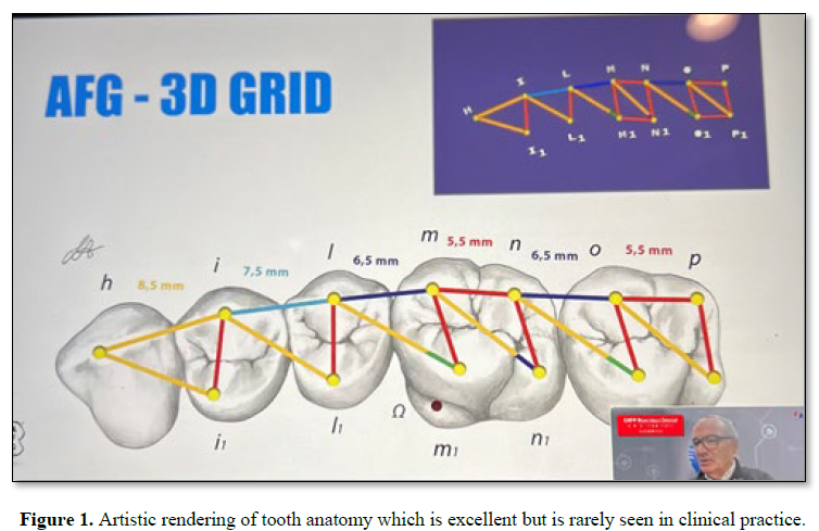
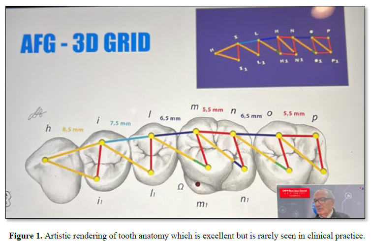
An occlusal contact results from two opposing teeth making contact upon closure. Contact areas can be big or small, intense or not intense. They are located using colored indicators [1]. A contact is a contact. An ideal contact has never been described. Just like a fingerprint occlusal contacts are individual and personalized. Contact areas are everywhere. How do we interpret them? What do they tell a practitioner? Contacts and associate areas may be bellwethers for health and disease. It all depends on how the clinician interprets what they see. We use papers and ribbons to record contacts [2]. They are hold overs from the past when typewriters became available in the late 1800’s. Ribbons and carbon papers were commonly used for typing and printing. Dentistry adapted them for contact marking. Early dentistry did not need indicators until the discipline of restorative dentistry was established. We still use them because they are easy to use and mark in red, black and blue. Clinicians interpret markings as they wish. They do not store them. They are taught to wash them away upon usage because they are only colored markings. There is no dental literature that describes a working contact. There are many descriptions of idealized contact points that are positioned on anatomically correct surfaces but they are misleading because in chair side dentistry surfaces are worn and there are no ideal contacts or contact locations [3] Figure. 1. Clinical dentistry is all about practicality.
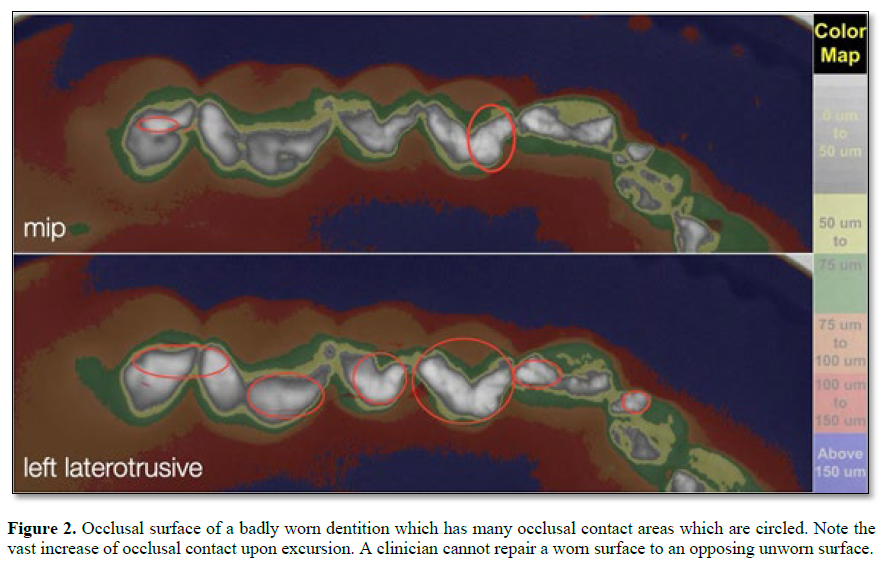
The figures demonstrate various means to monitor occlusal contact. How do we fit one contact into another to prevent occlusal damage. This is far reaching because it includes the working dentition and associated structures Figure. 2.
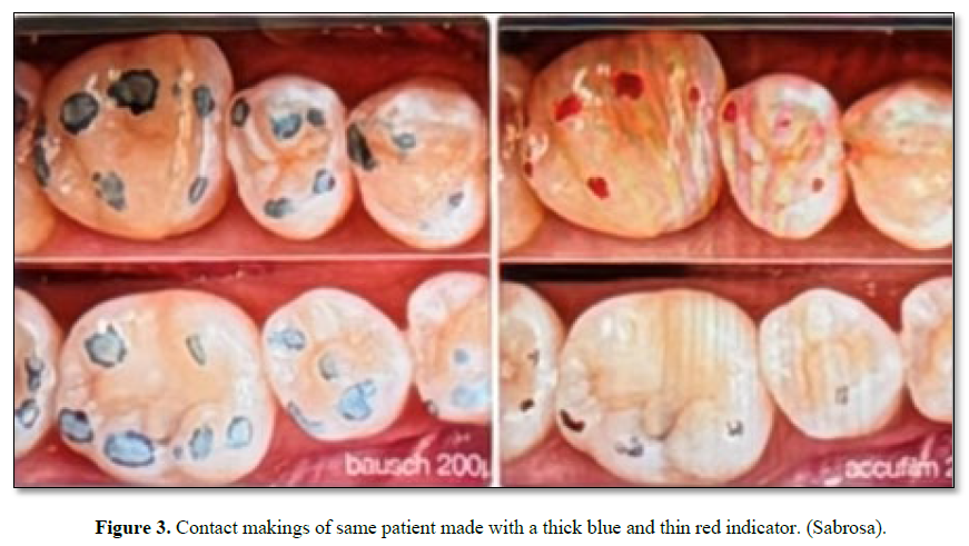
The only way a dentist can identify a contact is by marking it with an indicator. The indicator determines the mark. A 200 micron thick indicator makes many more markings than a 20 micron thin indicator Figure. 3.
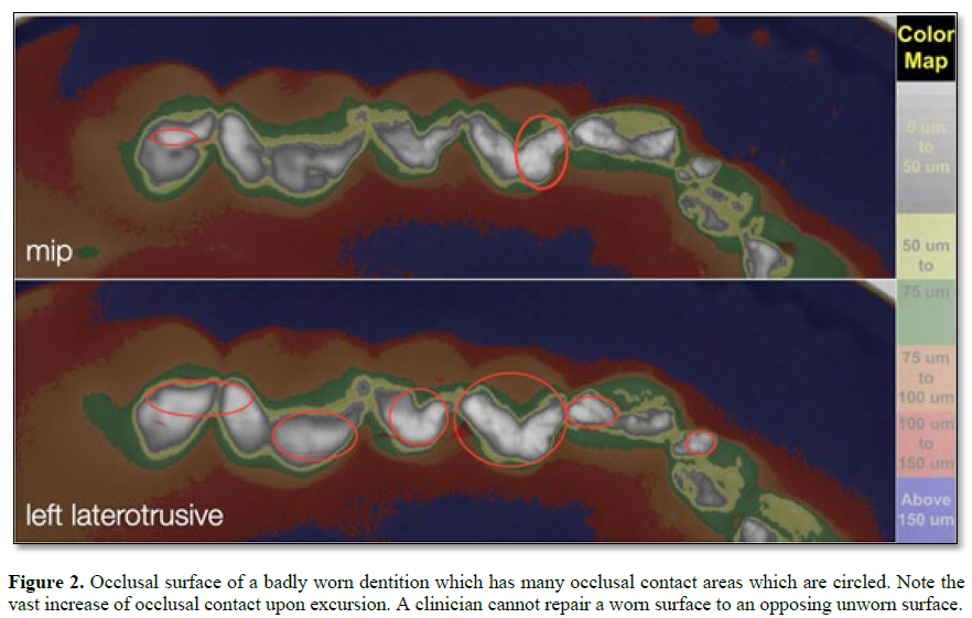
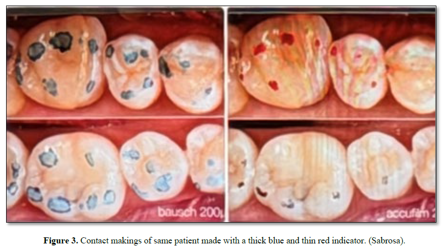
Clinically, tooth surface removal is dictated by contact markings. The bigger the mark the more the surface reduction. Surface removal is often made whether a mark is true or false. The thin indicator misses contacts and the thick indicator adds them. Contact markings tell a story but the clinician only sees a mark. Electronic devices do not suffice because they do not measure occlusal contact. They measure occlusal force from afar [4]. The only way to get a complete picture of occlusal contacts is to impress them and process the impression using image analysis. A clinician can then visualize the contact areas for size, location and intensity [5]. Permanent contact areas can also be compared to a baseline should one exist. Wear areas of contact and changes in intensity indicate positive or negative shifts in the occlusion [6]. Orthopedic surgeons evaluate bone density on radiographs by using image analysis [7]. Dental clinicians can also measure contact areas and intensities using image analysis [8]. The procedure that follows incorporates image analysis. It is fast and easily mastered. Take a bite impression using set or no set translucent silicone material in a triple tray. Place the impression on the surface of an illuminated light box and photograph it from above. The camera which is positioned above the illuminated impression is connected to an image analysis program (Image J). The resultant images are projected on an adjacent screen. The resultant black and white images are converted to colored images which represent contact and near contact areas [9]. Viewing is enhanced. Intense areas of contact can be numerically evaluated. This is foundational work because the clinician now has a permanent baseline to record wear and associated change. The anatomies of working contacts are revealed. Timely evaluation follows. Modified bite guard therapy becomes available [10]. Prevention is possible.

No Files Found


Internationally Accepted
Share Your Publication :